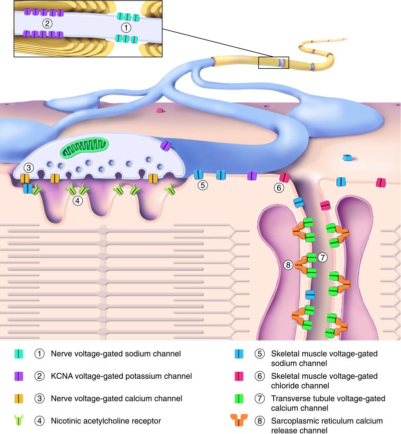
Figure 1.
Ion channels of the motor nerve and skeletal muscle involved in human disease. The drawing shows a myelinated axon branching to form synaptic contacts with a muscle fiber. (Upper) The outer surfaces of the axon and muscle. (Lower) The nerve presynaptic terminal and muscle fiber in cut section. (Inset) A magnified view of the nerve fiber, cut lengthwise near a node of Ranvier. Different channel types are identified by number and color, as indicated in the key below the drawing. The contractile proteins of the sarcomere are depicted schematically within the muscle. See text for additional details.

