
Figure 3.
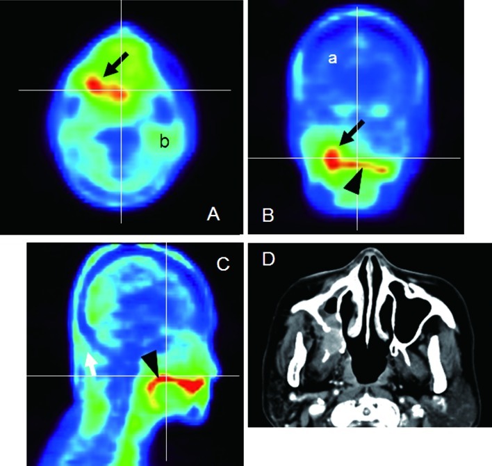
Mucoepidermoid carcinoma of the right maxilla (Case 2). (A) Axial, (B) coronal and (C) sagittal 18F-BPA-PET. The horizontal and vertical lines correlate to the cut surface of each image. As normal structures, the brain parenchyma (a) and parotid gland (b) were easily detected. The tumor was expressed as a high uptake area (arrows), which continued to the high uptake area that was the dorsum surface of the tongue (arrowheads). Differentiation between the two structures was difficult on the 18F-BPA-PET images. (D) Axial-enhanced CT.