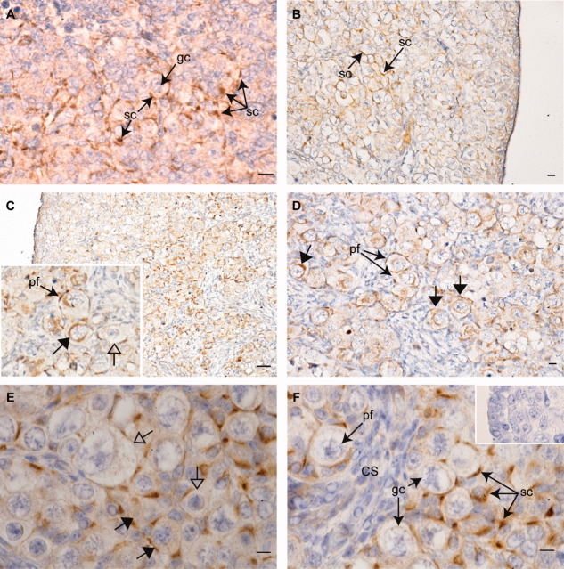
Fig. 2.
Immunohistochemical localization of brain-derived neurotrophic factor (BDNF) in the developing human fetal ovary. BDNF expression was detected at all stages examined. A: Expression of BDNF (brown staining) in somatic cells (sc) surrounding primordial germ cells (gc) in a first trimester (60 days gestation) human fetal ovary. B,C: At 14 (B) weeks and 20 weeks (C); BDNF is predominantly expressed in a corticomedullary gradient, with weak expression in somatic cells near the ovarian periphery, and intense expression in those interspersed between larger germ cells away from the periphery. C, inset: BDNF expression is detectable in the pregranulosa cells of primordial follicles (pf), and in some large germ cells (closed arrows), whereas others of comparable size and show no expression (open arrows). D: At 20 weeks; BDNF immunopositive germ cells are detectable within primordial follicles. E: At 18 weeks; BDNF expression varies between germ cells within the same nest, with immunopositive and immunonegative germ cells in existing close proximity. F: At 18 weeks; BDNF expression is strongest in somatic cells interspersed within germ cell nests and in primordial follicles. No expression is detectable in somatic cells within cell streams (cs). F, inset: negative control, primary antibody preincubated with immunizing peptide. Scale bars = 500 μm in A–D, 100 μm in E,F).

