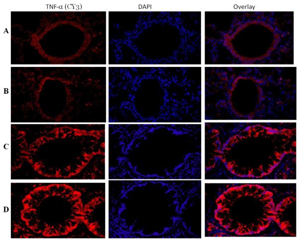
Figure 4. TNF-α expression in lung tissue.
Immunofluorescence of TNF-α expression in the lung of vitamin D-sufficient (C) and vitamin D-deficient (D) OVA-sensitized and challenged mice compared to PBS control vitamin D-sufficient (A) and vitamin D-deficient (B) OVA-sensitized and challenged mice, respectively (600× magnification). Sections were stained using rabbit anti-TNF-α antibody and goat anti-rabbit cy3 as secondary antibody. DAPI was used to stain the nuclei.

