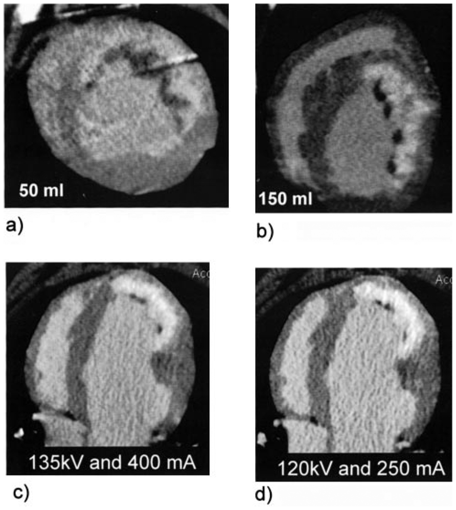
Figure 9.
Comparison of (a) low (50 mL) and (b) high dose (150 mL) MDCT infarct images 5 minutes after contrast injection. Panels c and d show identical slices acquired with 2 different imaging protocols to demonstrate the effect of radiation dose on image quality. The image in (c) was acquired with a relatively high radiation dose (135 kV and 400 mA, approximately equivalent to 3 REM), whereas the image in (b) was acquired with the clinical protocol used at our institution for CT coronary angiography (120 kV and 250 mA). Lower contrast and radiation does had no effect on the accuracy of quantitative infarct-size measurements.

