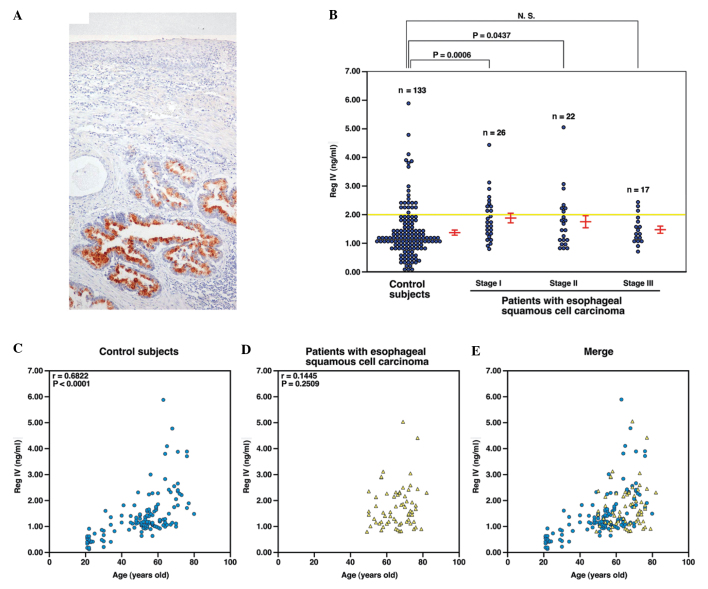
Figure 1.
Immunostaining and serum concentration of Reg IV in esophageal cancer. (A) Immunostaining of Reg IV in esophageal adenocarcinoma (original magnification, ×100). Reg IV staining is observed in goblet cell-like vesicles of adenocarcinoma cells. (B) Enzyme-linked immunosorbent assay of serum samples from 133 control subjects and 65 patients with esophageal squamous cell carcinoma (SCC). The yellow bar indicates the cut-off levels defined on the basis of a previous study (2 ng/ml) (15). The red bars indicate the mean ± SE. Differences in the serum concentration of Reg IV between two groups were tested using the Mann-Whitney U test. (C) Correlation between the serum concentrations of Reg IV and age in control subjects. The correlation was examined using Spearman’s rank correlation test. (D) Correlation between the serum concentrations of Reg IV and age in patients with esophageal SCC. The correlation was examined using Spearman’s rank correlation. (E) Panel (C) was merged with panel (D).

