Figure 4. .

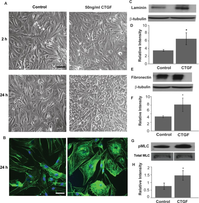
CTGF-induced changes in actin cytoskeletal organization, MLC phosphorylation, and ECM synthesis in HTM cells. Serum-starved HTM cells were treated with CTGF (50 ng/mL) or acetate buffer (control) for 24 hours and examined for changes in cell morphology, actin stress fibers, pMLC, and ECM proteins (fibronectin and laminin). (A) Phase contrast images revealed notable changes in cell morphology characterized by stiffened and contractile nature following treatment with CTGF after 24 hours compared to 2 hours of treatment and controls. This change in cell morphology was associated with increased actin stress fibers (phalloidin staining) in CTGF-treated TM cells (B). CTGF treatment for 24 hours also led to an increase in the levels of laminin (C, D) and fibronectin (E, F), and in the levels of phosphorylated MLC in HTM cells (G, H). Bars: 10 μm. β-tubulin and total MLC were probed as loading controls for ECM and phospho-MLC, respectively. *Denotes significant changes (P < 0.05, based on n = 4).
