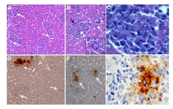
Figure 3.
Liver pathology and intracellular detection of Brucella antigens in macrophages of BALB/c mouse after 10 days of infection with virulent B. abortus 2308. (A) Liver granulomas (pointed by white arrows). (B) Large and smaller liver granulomas (white arrows) with giant cells (black arrow and insert). (C) Mononuclear infiltrate formed mainly by macrophages and histiocytes (white arrow). (D-E) immunoperoxidase detection of Brucella LPS antigen in matching histological sections of the corresponding upper A, B and C panels. Hematoxylin-eosin stain (A-C) and hematoxylin counterstain (D-F).

