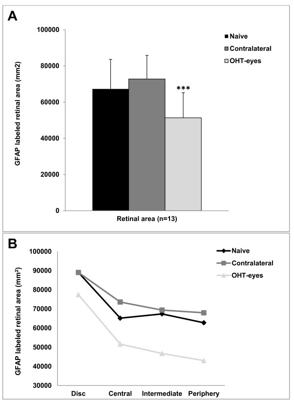
Figure 4.
GFAP-labeled retinal area (GFAP-RA) after 15 days of laser-induced OHT. Comparison among areas and concentric zones of the retina analyzed in the three study groups. The GFAP-RA of the OHT-eyes underwent a statistically significant reduction in comparison with naïve and contralateral eyes. This finding was observed when the analysis was made both by retinal areas (13 target areas per retina) (A) and by concentric zones of the retina (disc, central, intermediate, and periphery) (B). Each bar represents the mean ± SD of GFAP-RA. ***P < 0.001 versus naïve and contralateral retinas. ANOVA with Bonferroni test. ANOVA, analysis of variance; GFAP, glial fibrillary acidic protein; OHT, ocular hypertension.

