
Figure 7. Determinants of TGEV S antigenic site A.
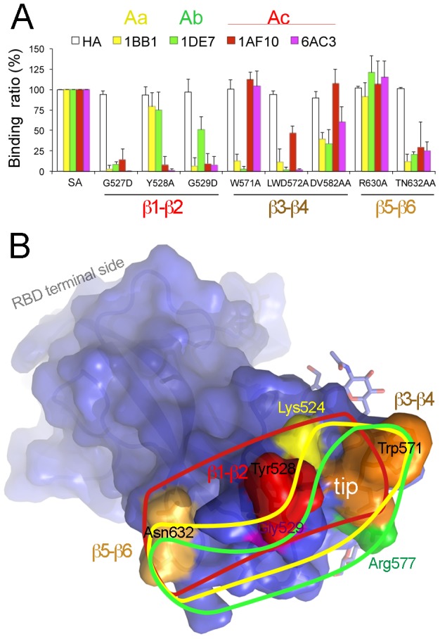
A. Binding of TGEV-neutralizing, site A-specific mAbs to RBD mutants. Relative binding (%) of mutants to wild type SA protein is shown for TGEV S-specific mAbs (top; described in Figure 1C) and a control anti-HA antibody (see Materials and Methods). RBD regions in which mutations locate are shown (bottom; see also Figure 5D). Mean and standard deviation of data from at least three experiments. B. Antigenic site A in the TGEV RBD and epitopes for antibodies. Surface and ribbon representation of the RBD with the 1AF10 contact regions colored as in Figure 2B. Three antibody-binding residues (Tyr528, Trp571 and Asn632) in the loops at the RBD tip, as well as TGEV Lys524, Arg577 and Gly529 residues associated with Aa, Ab and Ac subsites [24], respectively. Lines indicate epitopes for mAbs specific for each of the three antigenic subsites: Aa in yellow, Ab in green and Ac in red.