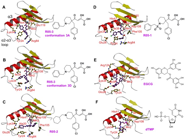
Figure 2. Binding of diketo inhibitors, EGCG and dTMP in the active site of pH1N1 endonuclease.
Manganese ions are pink spheres and the ion co-ordination is shown with green lines. Side chains of key active site residues that interact with the compound or are close to it are shown. The orientation in each case is the same after superposition of the domain. Helix α3 (red), the α3-α3 loop and beta strands β6, β7 and β8 (yellow) are indicated in panel A. A: R05-3 in conformation 3A. B: R05-3 in conformation 3D. C: R05-2 (chain A). Ala20 is marked in addition (see discussion). D: R05-1. E: EGCG. F: dTMP.

