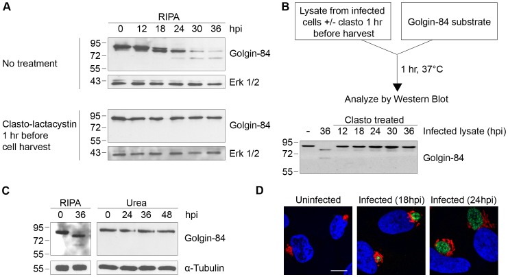
Figure 1. Golgin-84 cleavage does not occur in Chlamydia-infected cells when CPAF is inhibited during cell processing.
(A) Uninfected (0 hpi) and infected cells at time points between 12 and 36 hpi were treated with methyl acetate as a solvent control (top panel) or 150 µM of the CPAF inhibitor clasto-lactacystin (bottom panel) for 1 hour prior to cell lysis in RIPA buffer. Total cell lysates were separated by SDS-PAGE and probed with antibodies against golgin-84 or Erk 1/2 (loading control). (B) Cell-free degradation assay testing for CPAF activity in lysates prepared from the Chlamydia-infected HeLa cells described in Figure 1A. Each infected cell lysate was incubated with a lysate of uninfected HeLa cells as the source of golgin-84 substrate and reactions were analyzed by immunoblotting with golgin-84 antibodies. (C) Lysates of uninfected (0 hpi) or infected cells from different times in the infection were prepared in RIPA buffer (left panel) or by direct lysis in 8M urea (right panel), separated by SDS-PAGE and analyzed with antibodies to golgin-84 or α-tubulin (loading control). (D) Confocal images of uninfected or Chlamydia-infected HeLa cells examined at 18 and 24 hpi. Cells were stained with antibodies to the Golgi marker α-mannosidase II (red), the chlamydial major outer membrane protein MOMP (green) and the DNA dye Hoechst 33342 (blue) to detect Golgi membranes, the chlamydial inclusion and DNA, respectively. Scale bar, 10 µm.

