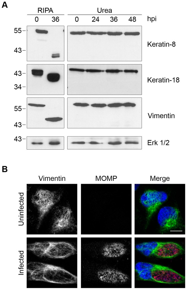
Figure 3. Cleavage of intermediate filaments in Chlamydia-infected cells is also dependent on cell processing.

(A) Lysates of uninfected (0 hpi) or infected HeLa cells were prepared in RIPA buffer (left panel) or by direct lysis in 8M urea (right panel) at the indicated times, separated by SDS-PAGE and analyzed by immunoblotting with antibodies to keratin-8, keratin-18, or vimentin. Equal loading for each blot was monitored by blotting for Erk 1/2 (loading control), but only the loading control for keratin-8 is shown. (B) Uninfected and infected HeLa cells at 30 hpi were fixed and stained with antibodies to vimentin (green), the chlamydial major outer membrane protein MOMP (red) and the DNA dye Hoechst 33342 (blue). Representative confocal images are shown. Scale bar, 10 µm.
