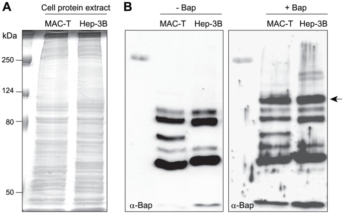
Figure 2. Identification of Gp96 as a Bap cellular receptor by a ligand overlayer assay.
A) Total protein extracts from MAC-T and Hep-3B cells were separated on a SDS gel, transferred to a nitrocellulose membrane by western-blotting and probed with/without pure Bap protein (50 µg/ml). B) Bound Bap protein was detected with polyclonal anti-Bap antibodies (α-Bap). A ∼100 kDa band reacting with anti-Bap antibodies is shown by the arrow. This protein was identified by MALDI-TOF analysis of the comigrating band on a Coomasie blue stained gel as Gp96.

