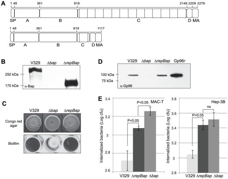
Figure 8. Effect of Bap repetitions in Gp96 interaction and cell invasion.
A) Structural organization of Bap with or without repetitions. SP, signal peptide; A, region A; B, region B; C, repetition region; D, region of serine-aspartate (SD) repeats; MA cell wall anchor. B) Western-blot analysis with anti-Bap (α-Bap) serum of surface protein preparations obtained under isosmotic conditions from S. aureus V329 (Bap+), Δbap and ΔrepBap (truncated Bap). C) Biofilm phenotypes of S. aureus V329, Δbap and ΔrepBap. Congo red morphology on congo red agar plates and biofilm formation in microtiter plates. D) Interaction of Bap-truncated protein with Gp96. S. aureus V329 (Bap+), Δbap and ΔrepBap strains were subcultured with 5 µg/ml of recombinant Gp96. Unbound Gp96 was removed by extensive washing and Gp96 bound to bacteria was detected using immunoblot analysis. E) Entry of S. aureus V329, Δbap and ΔrepBap strains was analyzed by gentamicin assay in MAC-T and Hep-3B cells. Experiments were performed in triplicate and repeated three times for each cell line.

