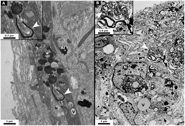
Figure 4. Micrograph of nerve cells of Porcellio d. dilatatus injected by wVulC.
These cells are localised in the CNS ganglion. (A) At 30 days post-injection, some autolysosomes and bacteria are visible: autophagic process is already quite high compared to control [N: Nucleus, V: Vacuole, Ap: Autophagosomes (Autophagic hallmark), Al: Autolysosomes, Wo: Wolbachia, *: a Wolbachia which seems to be surrounded by an autolysosome, Arrow head: magnified area]. (B) At 60 days post-injection, the nerve cells are severely damaged by the accumulation of autophagic vesicles (N: Nucleus, V: Vacuole, Ap: Autophagosomes (Autophagic hallmark), Al: Autolysosomes, Arrow head: magnified area and probably a Wolbachia in autophagic degradation).

