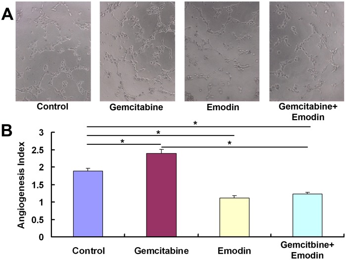
Figure 3. Effect of emodin on the angiogenesis of pancreatic cancer.
(A) Representative micrographs (200×) of angiogenesis after treatment of diluent control, gemcitabine (20 µmol/L), emodin (40 µmol/L), or their combination for 72 h. Cells were plated onto the Matrigel-precoated wells (5×10³ cells/well) and cultured in 10% FBS-DMEM medium. Original magnification. (B) Angiogenesis of endothelial cells (ECs) isolated from pancreatic cancer tissues. In vitro angiogenesis was quantitatively analyzed as described in Materials and Methods. *, P<0.05.

