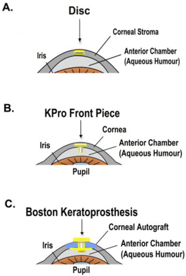
Figure 4.

Schematic diagrams of the various material models in the anterior eye of the rabbit. (A) The disc model shows the disc residing intrastromally in the cornea. (B) The KPro Front Piece (KPro-FP) model shows the front piece in the corneal stroma with its stem protruding into the anterior chamber. (C) The B-KPro model shows the PMMA front plate resting on the corneal epithelium with the PMMA stem extending through a corneal autograft, through a backplate, into the anterior chamber, and secured by a locking C-ring.
