
Figure 7.
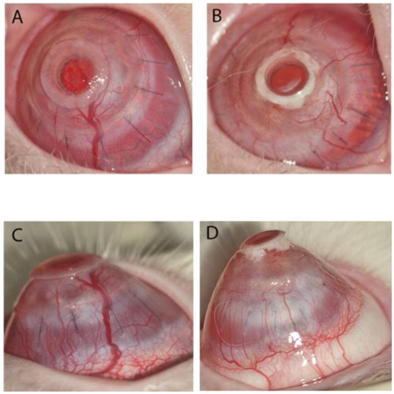
Clinical photographs of an anterior and profile view of comparative B-KPro Type I with a corneal autograft in the New Zealand white rabbit eye at 69 days post-operatively. (A, C) In the HMPEI-PMMA B-KPro rabbit eye, there is no mucous accumulation seen behind the front plate, but there is epithelial cell growth over the front plate, moderate neovascularization, and mild conical ectasia. (B, D) In the control PMMA B-KPro rabbit eye, there is visible accumulation of cellular debris, mucous in a space behind the front plate, moderate-severe corneal neovascularization, and corneal ectasia.