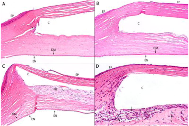
Figure 8.

Representative histopathology of a (A) HMPEI-Ti disc is shown compared to (B) control Ti disc at 61 days post-operatively. There is minimal cellular reactivity at the edges of the disc (arrow) and no significant difference between the two groups (hematoxylin and eosin stain; A ×100, B ×100). Histopathology shown for the HMPEI-PMMA KPro-FP (C) compared to control PMMA KPro-FP (D), at post-operative day 37. (C) HMPEI-PMMA KPro-FP shows minimal inflammatory response and a fibroblastic response is present posterior to the cavity (c) that contained the explanted KPro-FP. (D) The uncoated PMMA KPro-FP shows a moderate acute inflammatory response (arrows) surrounding the cavity (c). There is minimal microvascularization (V) (hematoxylin and eosin stain; C ×100, D ×200; DM, Descemet's membrane; EN, endothelium; EP, epithelium).
