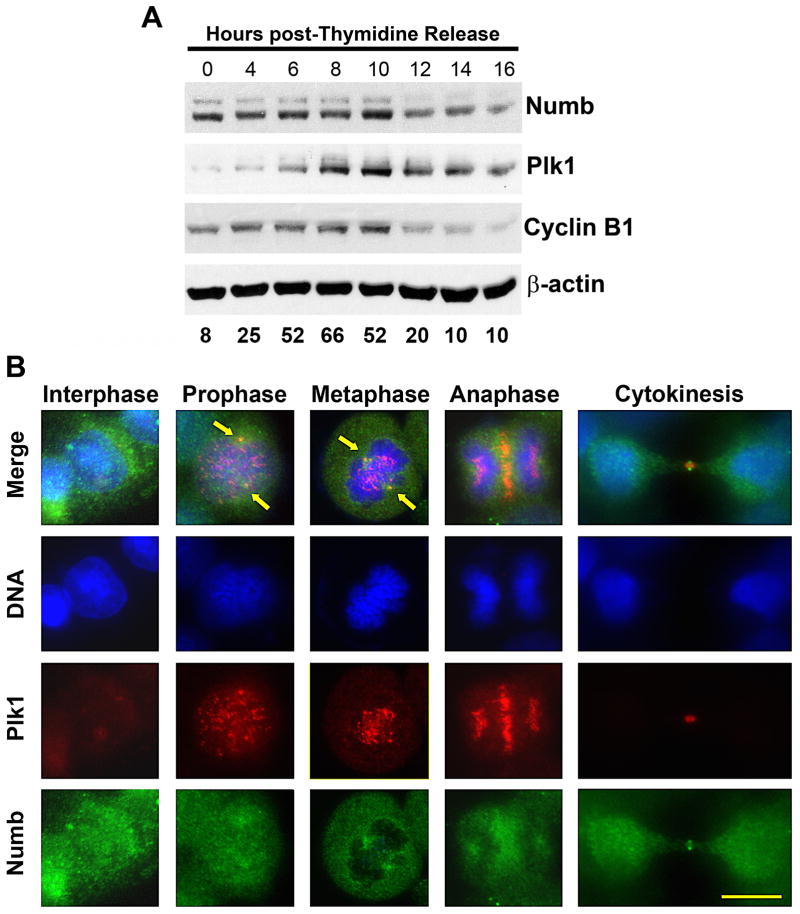
Figure 2. Numb expression is mitotically regulated.
A) Numb expression levels peak at mitosis, following a pattern similar to Plk1 and Cyclin B1. Using a double thymidine arrest and release protocol, A375 cells were collected for immunoblot and FACS analysis every two hours. Lysates were evaluated for Numb, Plk1 and Cyclin B1. Equal loading was confirmed by re-probing the blots for β-actin. The data represent three independent experiments where average G2/M percentage at each time point is shown. B) Plk1 and Numb co-localize during mitotic progression. To determine what role Numb may have during its peak at mitosis, the asynchronous A375 cells at various stages of mitosis were evaluated using immunofluorescence analysis. Cells were fixed and stained for Plk1 (red), Numb (green) and DNA (blue). Arrows indicate the co-localization at the spindle poles during prophase and metaphase. Scale bar = 10 μm.

