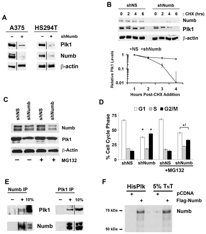
Figure 3. Numb regulates Plk1 protein levels through direct interaction.
A) Numb knockdown decreases Plk1 expression. A375 and HS294T cells were treated with nonsense (NS), Plk1 or Numb targeting shRNA, lysates were collected and evaluated via immunoblot analysis for Plk1 and Numb. Equal loading was confirmed by re-probing the blots for β-actin. Results shown are from the same membrane, line denotes removal of a single lane between the two samples. B) Numb is required for Plk1 protein stability. To evaluate if Numb positively regulates Plk1 protein half-life, A375 cells were treated with nonsense (NS) or Numb targeting shRNA then with cycloheximide (CHX) to inhibit new protein synthesis. Following treatment with CHX (0, 2, 4 and 6 hours), lysates were collected and analyzed by immunoblot analysis for Plk1 and Numb. Equal loading was confirmed by re-probing the blots for β-actin. The protein levels were quantitated by a densitometric analysis of protein bands. The data (relative density normalized to β-actin) is expressed as mean ± standard deviation of three experiments on a log10 scale. C) Numb stabilizes Plk1 protein by blocking proteasomal degradation. A375 cells were treated with nonsense (NS) or Numb targeting shRNA in the presence or absence of 10 μM MG132 to block proteasome mediated degradation for 8 hours. Following treatment, lysates were collected and analyzed by immunoblot analysis for Plk1, Numb and β-actin as a loading control. D) Proteasome inhibition normalizes the cell cycle abnormalities of Numb knockdown. After treatments described in Figure 2C, cells were collected and analyzed for cell cycle profile using FACS analysis. Data represents mean ± standard deviation of three separate experiments with similar results (*p<0.01 relative to shNS within respective vehicle or MG132 treatment, !p<0.01 relative to shNumb of vehicle treated cells). E) Endogenous Plk1 and Numb co-immunoprecipitate in vivo. Cell lysates were prepared from actively dividing A375 cells and immunoprecipitations were performed with Plk1, Numb or their respective IgG control antibodies. The immunoprecipitates were separated by SDS-PAGE with a 10% aliquot of untreated lysate and probed for the converse target using immunoblot analysis. “−” denotes addition of animal specific IgG inclusion as control, “+” denotes antibody included in immunoprecipitation. Results shown here are from same membrane, the line denotes the removal of extraneous lanes. F) Plk1 and Numb directly interact in vitro. To confirm if the interaction between Plk1 and Numb is a direct interaction or requires a third priming kinase or mediator we in vitro translated and transcribed Flag-Numb plus [35S]-Methionine using Promega’s TNT system. The TNT product was mixed with His-Tagged recombinant Plk1 and purified using Ni-Magne-His purification system. Purified lysates were separated using SDS- PAGE along with 5% of the TNT reactions, dried and analyzed by autoradiography.

