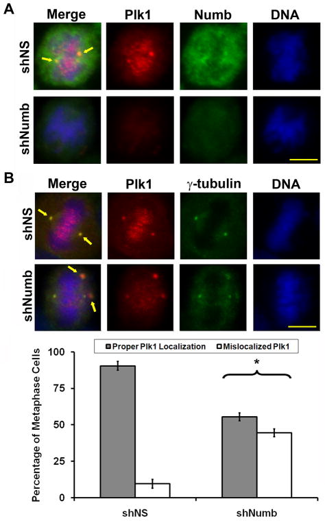
Figure 4. Numb regulates the localization of Plk1 during metaphase.
A) Plk1 localization is dysregulated after Numb knockdown. A375 melanoma cells were treated with nonsense (NS) or Numb targeting shRNA then fixed and stained for Plk1 (red), Numb (green) or DNA (blue). Representative metaphase images from each treatment are shown. Arrows indicate the typical overlap of Plk1 and Numb staining. B) Mislocalization of Plk1 during Numb knockdown results in disorganized centrosomal γ-tubulin recruitment. To evaluate if the mislocalized Plk1 after Numb knockdown is a result of a multipolar phenotype, A375 melanoma cells were treated with nonsense (NS) or Numb targeting shRNA, fixed and stained for Plk1 (red), γ-tubulin (green), or DNA (blue). Because Numb knockdown results in decreased Plk1 staining intensity the Red channel (Plk1) was increased by 50% to visualize mislocalized Plk1. At least 100 metaphase cells in three independent trials (non mono-polar) were counted and scored as possessing proper Plk1 localization (aligned at the centrosomes with γ-tubulin) or mislocalized Plk1 (increased punctate signal off the centrosome or metaphase plate). Data represents the mean ± standard deviation of three independent trials (*p<0.01). Arrows indicate normal Plk1 and γ-tubulin overlap in first row and mislocalized Plk1 away from the centrosome and/or off the metaphase plate in the second row. Scale bar = 10 μm.

