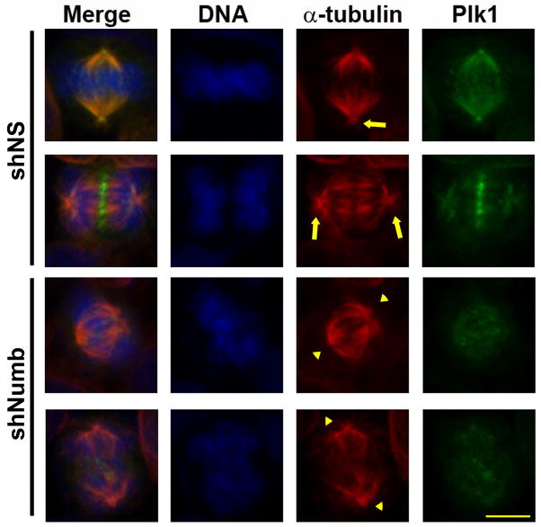
Figure 5. Reduced γ-tubulin recruitment results in loss of mitotic aster formation.
To determine if loss of Numb affects α-tubulin dynamics A375 melanoma cells were treated with nonsense (NS) or Numb targeting shRNA, fixed and stained for Plk1 (green), α-tubulin (red), or DNA (blue). Because Numb knockdown results in decreased Plk1 staining intensity the Green channel (Plk1) was increased by 50% to visualize mislocalized Plk1. Arrows indicate presence of aster microtubules and arrowheads highlight the lack of aster microtubules. Scale bar = 10μM.

