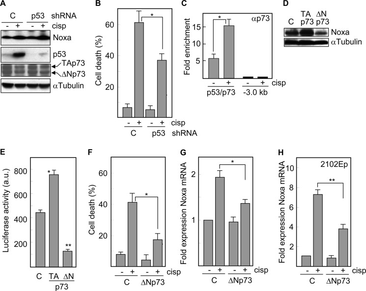
FIGURE 5.
Cisplatin-induced expression of Noxa is dependent on p73 but not p53 in embryonal carcinoma cells. NTERA2 cells transfected with a vector expressing a shRNA targeted against p53 were treated with cisplatin and A, the expression levels of Noxa, p53 and p73 isoforms were determined by Western blot analysis, and B, cell death was determined following 24 h of treatment. C, ChIP analysis of the association of p73 with the Noxa promoter in the presence or in the absence of cisplatin. Immunoprecipitates were analyzed by quantitative PCR using primers specific to the target site or a 3 kb upstream region. Fold enrichment is expressed relative to IgG immunoprecipitates. D, protein levels of Noxa were analyzed in NTERA2 cells overexpressing an active (TA) or a dominant negative form (ΔN) of p73. Samples were run on the same gel but not in consecutive lanes. The levels of α-tubulin were also analyzed to assure equal loading. E, p53 knockdown cells were transfected with TAp73 or ΔNp73 and a luciferase-Noxa promoter vector and then analyzed for luciferase activity. F, p53 knockdown cells transfected with ΔNp73 were treated with cisplatin and 24 h later cell death was quantified. G, p53 knockdown NTERA, and H, 2102EP cells were transfected with ΔNp73 and treated with cisplatin for 24 h. The mRNA levels of Noxa were determined by quantitative RT-PCR. *, p < 0.01; **, p < 0.001. Histograms represent the mean ± S.D. of three independent experiments.

