FIGURE 4.
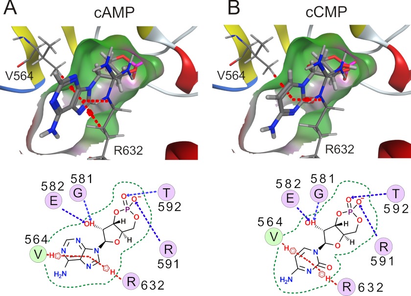
cCMP binds to CNBD of HCN2 channels. Structural models of a portion of the CNBD of HCN2 with bound cAMP (A) and cCMP (B) are shown (upper). The pocket is visualized by a molecular surface (hydrophobic (green) and hydrogen bonding (magenta)). Interactions between the protein and cyclic nucleotide are shown schematically (lower). Dashed lines indicate π stacking (red) and hydrogen bonding (blue). The border of the binding pocket is marked by dashed green lines.

