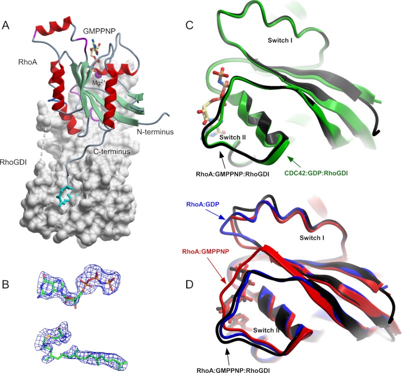
FIGURE 6.
Structure of RhoA·GMPPNP-GG·RhoGDI complex (PDB 4F38). A, overall structure of the complex. RhoGDI is displayed as a gray molecular surface, whereas RhoA is displayed in ribbon representation. The nucleotide and the conjugated geranylgeranyl isoprenoid are displayed in ball-and-stick representation. The Mg2+ is displayed as a space-filling magenta ball. B, 2.5 σ (Fo − Fc) difference in electron density of the bound GMPPNP and geranylgeranylated cysteine at the RhoA C terminus before incorporation into the model. C, superimposition of the prenylated CDC42·GDP and RhoA·GPPNHP complexed with RhoGDI. D, as in C but RhoA·GMPPNP complexed with RhoGDI is superimposed with unbound GMPPNP-associated (red) and GDP-associated (blue) forms of RhoA.

