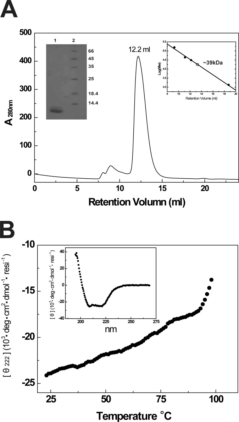
FIGURE 2.
Assembly and biophysical characterizations of the 6-HB formed by gp41 NHR546–588/CP32M chimera. A, size-exclusion chromatography and SDS-PAGE analyses of HIV gp41 NHR546–588/CP32M chimera. Chromatographic profile shows UV absorbance at 280 nm. Upper right insert, the log (molecular weight) values of the standard proteins for size-exclusion column calibration (Superdex 75 10/300 GL) are plotted as the function of Ve (elution volume) (●). The data are fitted linearly to derive the standard curve. The molecular mass of HIV gp41 NHR546–588/CP32M chimera is calculated as∼39 kDa (○). Upper left insert, SDS-PAGE analysis of purified HIV gp41 NHR546–588/CP32M chimera. Lane 1, HIV gp41 NHR546–588/CP32M chimera (∼12kDa); lane 2, molecular weight standards. B, the melting curve of NHR546–588/CP32M chimera showing the thermostability of NHR546–588/CP32M chimera. Insert, normalized circular dichroism spectrum showing the α-helical conformation of the 6-HB formed by the chimera. resi, the number of the residues.

