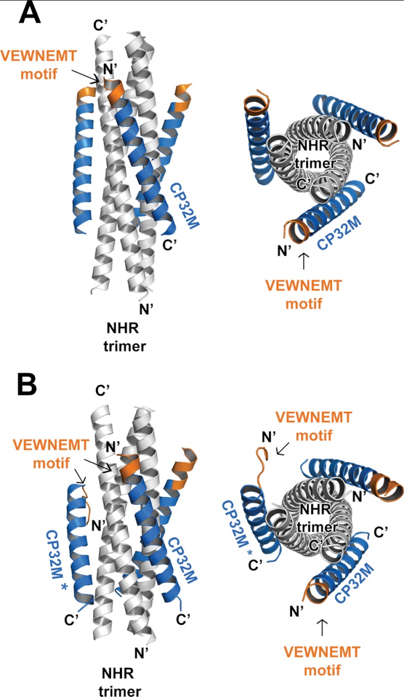
FIGURE 3.
Overall structure of the 6-HB structures formed by HIV gp41 NHR546–588/CP32M chimera. Ribbon models of two 6-HB structures formed by NHR546–588/CP32M chimera. The NHR trimers are colored in gray, the CP32M peptides are colored in blue, and the VEWNEMT motifs are colored in orange and labeled. The same color scheme as for the 6-HB is used in all of the figures that follow (Figs. 4–7). The CP32M molecule with the disordered N-terminal motif is marked with an asterisk. A, crystal form 1 (space group P321). Left, side view; right, top view. B, crystal form 2 (space group P21). Left, side view; right, top view.

