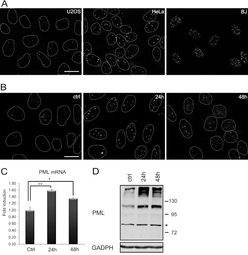
FIGURE 1.
PML and PML NBs are controlled by paracrine signaling. A, immunofluorescence detection of PML NBs in untreated U2OS, HeLa, and BJ cells cultured under standard conditions is shown. Immunofluorescence detection of PML NBs (B) and mRNA levels of PML (C) were quantified by qRT-PCR in U2OS cells treated 24 and 48 h with conditioned medium from BJ cells (diluted 1:1 with fresh medium). The values represent the average of two independent experiments performed in triplicate and are given as -fold induction of PML mRNA levels relative to control U2OS cells treated with conditioned medium from U2OS cells (diluted 1:1 with fresh medium); error bars represent S.E. Asterisks (*) and (**) represent p values <0.05 and <0.01, respectively. β-Actin was used as a reference gene. Bar, 15 μm. D, shown is immunoblot detection of PML in U2OS cells after 24 and 48 h of treatment with conditioned medium from BJ cells (diluted 1:1 with fresh medium). Bands with lower mobility than 130 kDa represent unmodified PML isoforms. A nonspecific band is marked by asterisk. GAPDH was used as a loading control.

