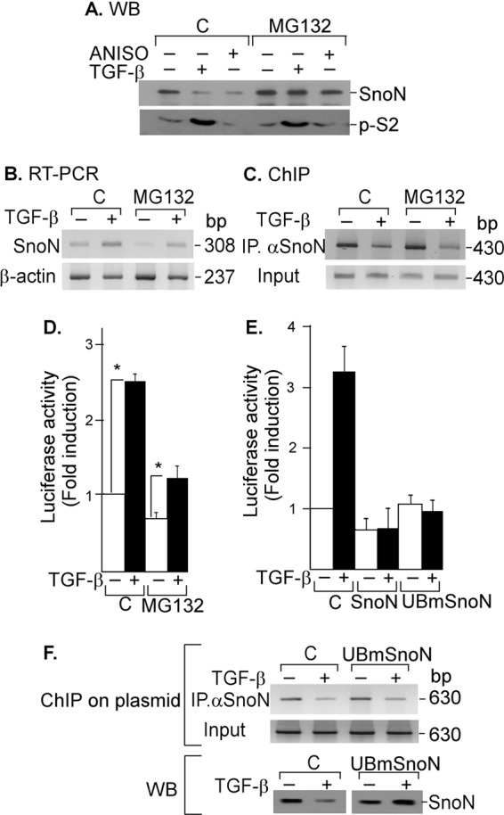
FIGURE 4.

TGF-β signal removes SNON from SKIL gene promoter independently of its degradation. A, A549 cells were pretreated for 2 h without or with 50 μm MG132 and then incubated for 45 min in the absence or presence of 300 pm TGF-β or 10 μm ANISO. Proteins were immunoprecipitated with anti-SNON or anti-SMAD2 antibody followed by WB (n = 2) (A), or total RNA was isolated, and SNON (308-bp) and β-actin (237-bp) mRNAs were amplified by RT-PCR with specific primers (n = 2) (B), or ChIP assays were carried out using anti-SNON antibody, and PCRs were done with primers spanning SKIL SBE region (430 bp) (n = 3) (C). D, A549 cells transfected with the skilSBEs(408)-Luc reporter were pretreated for 2 h without or with 50 μm MG132 and then incubated for 12 h with or without 100 pm TGF-β. Luciferase activity was evaluated and normalized using β-gal expression and is reported as -fold induction over control. Values are mean ± S.E. (error bars) of three separate experiments in triplicate. *, p < 0.05 compared with control (C). E, to further analyze whether SNON degradation was required to regulate SKIL gene, we used UBmutSNON, which is unable to be ubiquitinated or degraded. AD293 cells were transiently transfected with skilSBEs(408)-Luc reporter along with WT SNON or UBmSNON, and 24 h post-transfection, cells were incubated for 12 h with or without 100 pm TGF-β. Luciferase activity was evaluated and normalized using β-gal expression and is reported as -fold induction over control. Values are mean ± S.E. (error bars) of three separate experiments in triplicate. F, AD293 cells were transiently transfected with the skilSBEs(408)-Luc reporter with or without UBmSNON. Cells were incubated for 45 min with or without 500 pm TGF-β, and a ChIP on plasmid assay was carried out using anti-SNON antibody for IP. PCRs were done with primers spanning the SBE region cloned into pGL3 vector (630 bp) (upper panel). Endogenous SNON and UBmSNON protein levels were detected by Western blot (lower panel).
