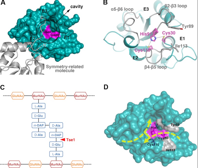
FIGURE 3.
Substrate-binding sites of Tse1. A, surface representation presenting the substrate-binding sites of Tse1. The catalytic Cys-30 and His-91 are colored in pink. The cavity is highlighted, and the entrances are labeled. The symmetry-related molecule is shown as a ribbon diagram. B, ribbon diagram of the substrate-binding sites of Tse1. The three protruding loops surrounding the cavity are labeled. The catalytic residues are shown as stick models and colored in pink. Tyr-89 and Ile-113, which clamp the scissile bond for hydrolysis, are also shown as stick models. C, schematic view of the cross-linked cell wall murein substrate. The scissile bond cleaved by Tse1 is highlighted. D, surface view showing the possible binding fashion of the cross-linked murein peptide substrate.

