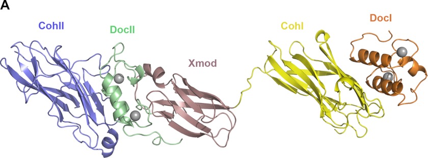
FIGURE 1.
Crystal structure of the DocI·CohI9–X-DocII·CohII ternary cellulosomal complex. One representative molecule of the DocI· CohI9–X-DocII· CohII ternary complex crystal structure is shown. The backbone ribbon representation depicts SdbA CohII in blue, the CipA DocII in green, X module in rose, CohI9 in yellow, and the Cel9D DocI in orange. Calcium ions are shown as gray spheres.

