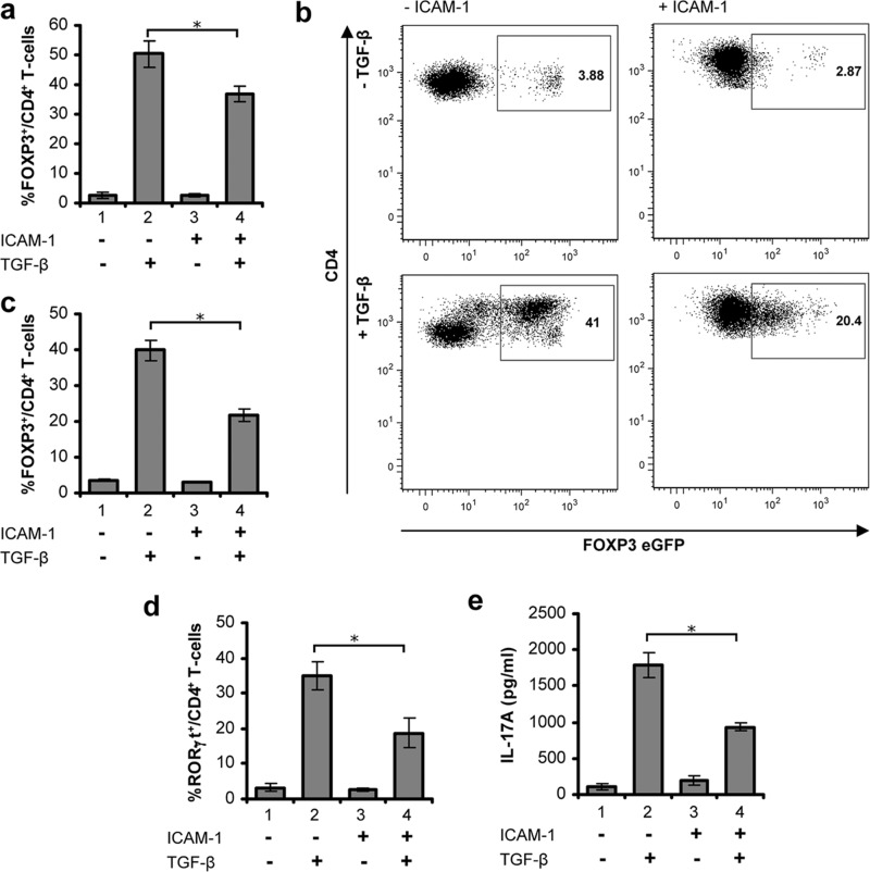
FIGURE 6.
LFA-1/ICAM-1-mediated signaling in T-cells suppresses TGF-β-induced FOXP3+ iTreg or RORγt+ Th17 differentiation. a, unstimulated or LFA-1/ICAM-1 stimulated human PBL T-cells were cultured with or without 5 ng/ml TGF-β under iTreg differentiation conditions for 5 days. Cells were stained for CD4 and FOXP3 followed by high content imaging and analysis. Percentage of FOXP3+ cells among CD4+ T-cells are presented. b, representative flow cytometry plots of Foxp3-eGFP expressing CD4+ T-cells from FOXP3-eGFP reporter mice. CD4+ cells were isolated and incubated with or without ICAM-1 under iTreg conditions for 72 h in the presence or absence of 5 ng/ml TGF-β. c, mean percentage of FOXP3-eGFP+ mouse CD4+ cells treated as indicated (data are mean ± S.E. from four to six individual mice). d, unstimulated or LFA-1/ICAM-1-stimulated human PBL T-cells were cultured with or without 5 ng/ml TGF-β under Th17 conditions for 4 days. Cells were stained for CD4 and RORγt, followed by high content imaging and analysis. Percentage of RORγt+ cells among CD4+ T-cells is presented. e, expression of IL-17, detected by ELISA, in supernatants from cells cultured as described in d. Data are representative of at least three independent experiments (mean ± S.E.). *, p < 0.05.

