Figure 1.
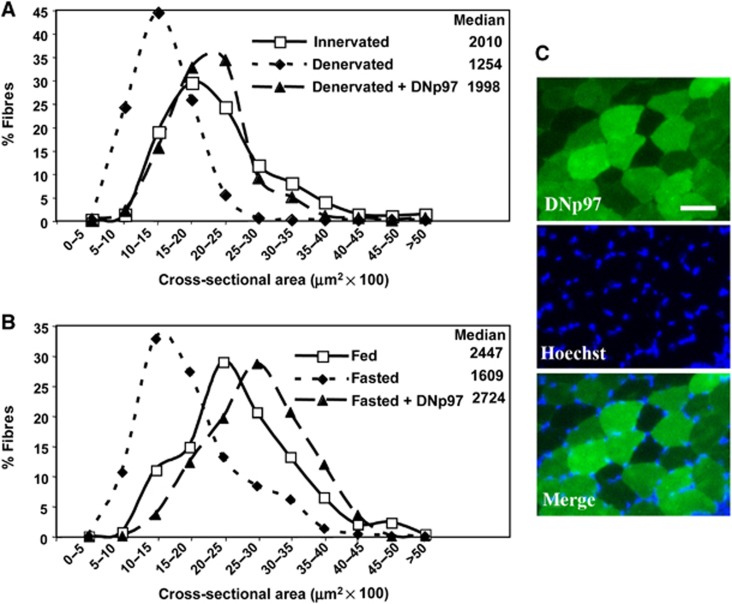
Electroporation of DNp97 in TA blocks both denervation and starvation-induced atrophy. (A) Frequency histograms showing the distribution of cross-sectional areas of muscle fibres of TA either innervated or 9 days denervated and transfected or not with DNp97GFP. Muscles were electroporated with DNp97GFP plasmids at the same time as section of the sciatic nerve. According to the Kruskal–Wallis Test, differences (P<0.001) were found between innervated versus denervated and innervated versus denervated+DNp97. (B) Frequency histograms showing the distribution of cross-sectional areas of muscle fibres of TA either from fed or 2 days fasted mice and transfected or not with DNp97GFP. Muscles were electroporated with DNp97GFP plasmids and after 4 days mice were deprived of food for 2 days. According to the Kruskal–Wallis Test, differences (P<0.0001) were found between fed versus fasted, fed versus fasted+DNp97 and fasted versus fasted+DNp97. (C) A representative field of a transverse section of fibres expressing DNp97GFP from food-deprived mice. Scale bar represents 50 μm.

