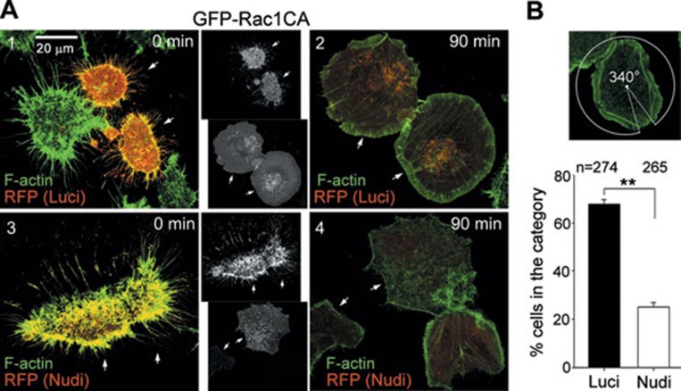
Figure 7.
Depletion of Nudel impairs the formation of the lamellipodial F-actin. (A) Nudel-depleted cells lacked branched actin network at cell periphery during re-spreading. GFP-Rac1CA-positive ECV304 cells transfected with pTER-Luci-RFP or pTER-Nudi-RFP were treated with EDTA for 5 min and then allowed to re-spread prior to fixation at the indicated time. F-actin was visualized with Alexa Fluor 647-conjugated phalloidin. The arrows indicate the cells positive for both GFP and RFP. (B) The statistical results for cells with the lamellipodia coverage ≥ 270° at 90 min. The coverage was measured as the size of the circumference angle that covered the entire lamellipodia as demonstrated. **P ≤ 0.01 in Student's t-test.

