
Figure 2. Defining structures of the cardiac loop.
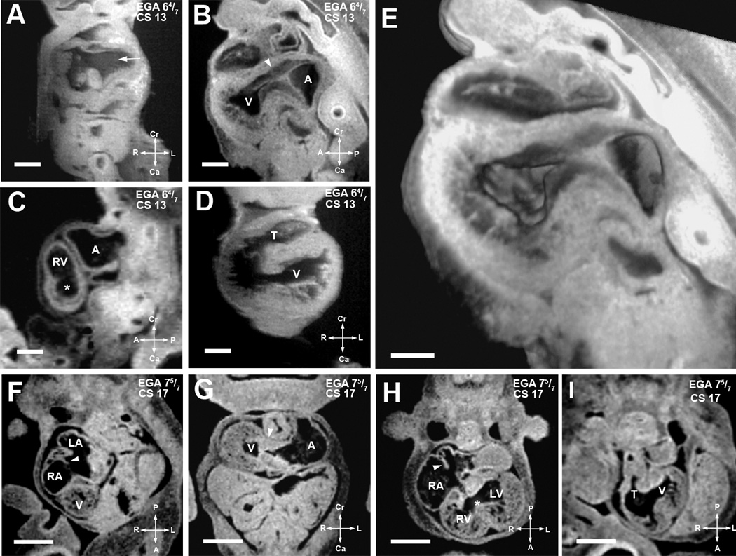
(A–E). EFIC and MRI images of EGA 6 4/7 weeks (CS13) embryos shown in various imaging planes. Imaging in the frontal plane (A) shows the common cardinal veins or the open venous confluence (arrow), while sagittal view (B) shows primitive endocardial cushions at the atrioventricular junction (arrowhead). A 3D model of the same embryo (E) shows the extent of the interventricular foramen as well as the contour of the endocardial cushions. MRI image of another embryo in the sagittal plane (C) shows the presumptive right ventricle (RV), atrial chamber (A), and a nondistinct interventricular foramen (*), while the ventricular chamber (V) and a single, undivided truncus arteriosus (T) can be seen in an frontal section of a third embryo (D).
Scale bars: (A–D)=0.4 mm, (E)=0.25 mm
(F–I). MRI images of EGA 7 5/7 weeks (CS17) embryo. Image from an oblique transverse plane (F) shows the right and left atrial (RA, LA) chambers as septation is progressing (arrowhead). The developing ventricle (V) is seen. Viewed in the transverse plane in (G), well formed dense endocardial cushion tissue is seen at the atrioventricular junction (arrowhead). Another section in the transverse plane in (H) shows the right and left ventricular cavities with a more distinct interventricular foramen (*). Septum primum can be seen as atrial septation progresses (arrowhead). The single undivided truncus arteriosus (T) and interventricular foramen communicating with the presumptive left ventricle (V) can be seen in an oblique transverse plane (I). Scale bar: (E–H) = 1.250 mm.
LV: presumptive left ventricular chamber, T: truncus arteriosus.