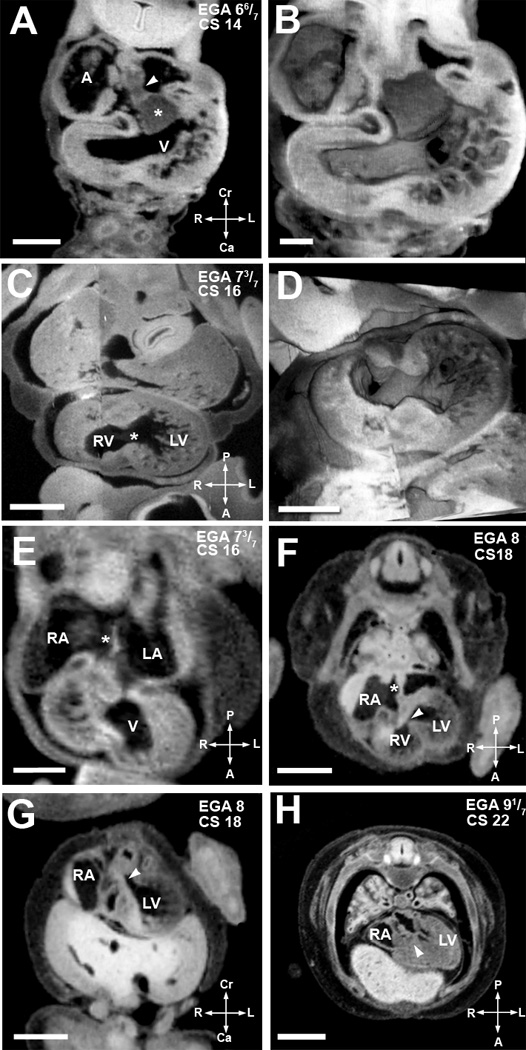
Figure 3. Major events of atrial and ventricular septation.

(A,B). EFIC image of EGA 6 6/7 weeks (CS14) embryo in the transverse plane (A) shows the atrial spine (arrowhead) attached to the inferior cushion (asterisks). 3D reconstruction (B) highlights the endocardial cushions and trabeculation in the ventricular chamber. Scale bar = 0.515 mm in (A), 0.272mm in (B).
(C,D). An EFIC image of EGA 7 3/7 weeks (CS16) embryo in the oblique plane (C) showing right and left ventricular chambers connected by an interventricular foramen (*). 3D reconstruction of the same embryo (D) delineates the contour of the interventricular foramen and the orifices of the atrioventricular canal and the truncus arteriosus. Scale bar = 0.5mm for (C), and 0.900mm for (D).
(E) MRI image of an embryo at EGA 7 3/7 weeks (CS16) also in the transverse plane. It shows the formation of septum primum (*) between the right and left atria (RA, LA). Scale bar = 0.5 mm.
(F,G) MRI image of an EGA 8 weeks (CS18) embryo in an oblique transverse plane (F) shows a complete atrial septum (*). The most caudal portion of the septum primum, the mesenchymal cap, has fused to the superior cushion. The growth of the muscular ventricular septum into the ventricular cavity is also shown. The crest of the muscular interventricular septum is present with an incomplete inlet ventricular septum (arrowhead) immediately above it. Panel G, another MRI image of the same embryo in an oblique coronal plane, shows the formed outlet ventricular septum (arrowhead). Together these two images show that outlet ventricular septation is completed before inlet ventricular septation. Scale bars in (F,G) = 1.5 mm.
(H). MRI image of embryo at EGA 9 1/7 weeks (CS22) in an oblique coronal plane shows a completed inlet ventricular septum (arrowhead). Scale bar = 2mm.
A: primitive atrium/venous confluence, RA: right atrium, LA: left atrium, V: ventricular chamber, LV: left ventricular chamber, RV: right ventricular chamber.
