
Figure 4. Major milestones of atrioventricular valve morphogenesis.
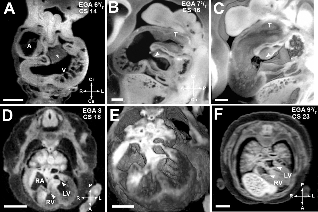
(A). EFIC image of an embryo at EGA 6 6/7 weeks (CS 14) in the transverse plane shows a large endocardial cushion (*) in the center of the cardiac loop. Scale bar = 0.515 mm
(B,C). EFIC image of an embryo at EGA 7 3/7 weeks (CS16) in a sagittal plane (B) shows a tight, well formed atrioventricular junction (arrowhead) and the truncus arteriosus (T). 3D volume of the same embryo (C) shows exquisite detail of the contour and shape of the endocardial cushions and the truncus arteriosus (T). Scale bar in (B,C)=0.389mm.
(D,E). MRI image in an oblique transverse plane (D) of an embryo at EGA 8 weeks (CS 18) shows separate atrioventricular valves. The valve leaflets appear thick at this stage. Note right and left atrioventricular valves denoted by arrowheads. 3D volume of the same embryo (E) shows indentation associated with the opening in the inlet ventricular septum. Scale bar for (D)=1.5 mm, (E)=1.1mm.
(F) MRI image in an oblique transverse plane of a more mature EGA 9 3/7 weeks (CS 23) embryo. It shows separate atrioventricular valves with thinner valve leaflets (arrowheads). The inlet septum is closed. Scale bar = 2 mm.
A: primitive atrium, RA: right atrium, V: ventricular chamber, RV: right ventricular chamber, LV: left ventricular chamber