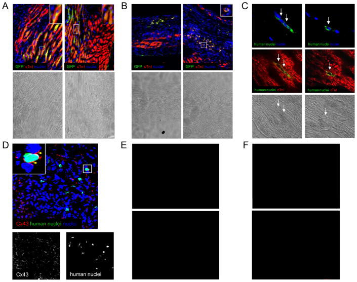
Figure 1.
Cardiogenic differentiation of transplanted cells cultured from humans biopsies using the cardiosphere method. Human CDCs were injected into SCID mice at the time of myocardial infarction. (A): Example images of the infarct border zone demonstrating engraftment and differentiation of lentiviral GFP-labelled CDCs 6 weeks after injection. (B): Example images of the central scar demonstrating rounded GFP-labelled CDCs weakly expressing markers of cardiac differentiation six weeks after injection. (C) Examples of unlabeled human CDCs within the infarct (left image) and border zone (right image) 3 weeks after injection. (D) Example of human-derived cardiomyocytes within the infarct border zone expressing Cx43 one week post-transplantation. (E) Example image of beta-galactosidase-labelled human CDCs differentiating into smooth muscle cells 3 weeks after injection. (F) Example image of beta-galactosidase-labelled human CDCs differentiating into endothelial cells 3 weeks after injection.

