Fig. 3.
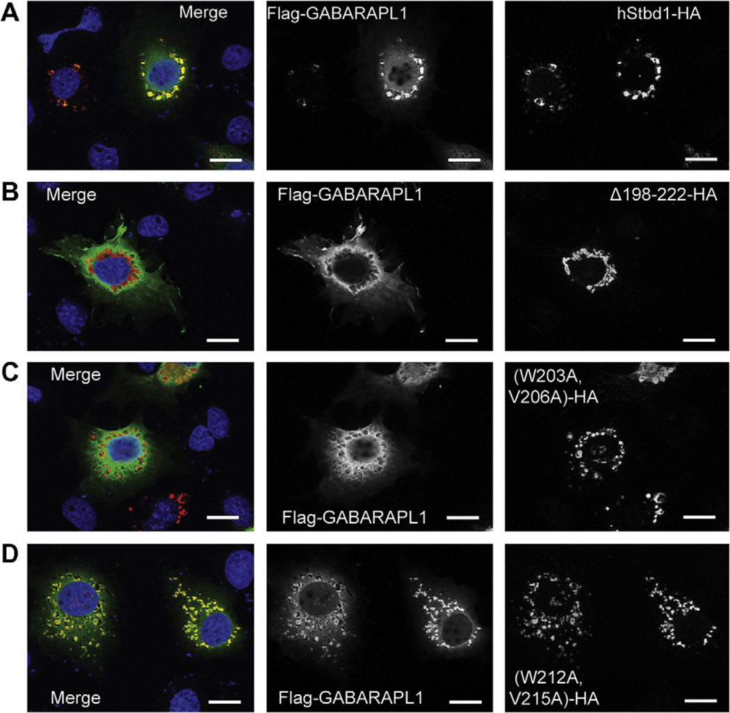
Subcellular localization of GABARAPL1 and Atg8 family interacting motif (AIM) mutants of Stbd1 co-expressed in COS M9 cells. Mutated hStbd1 with a C-terminal HA-tag was co-expressed in COS M9 cells with N-terminal Flag-tagged GABARAPL1 and immunostained with anti-HA antibodies (red) or anti-Flag antibodies (green). (A) Co-localization of hStbd1 and GABARAPL1 (merged in left panel) in cells co-expressing C-terminal HA-tagged full length hStbd1 (right panel) and N-terminal Flag-tagged GABARAPL1 (middle panel). (B) Loss of co-localization (merged in left panel) of Flag-tagged GABARAPL1 (middle panel) with potential AIM deletion mutant of hStbd1, Δ198–222–HA (right panel). (C) Impaired co-localization (merged in left panel) of Flag-tagged GABARAPL1 (middle panel) with double mutation in a potential AIM on hStbd1, (W203A, V206A)-HA (right panel). (D) Unaffected co-localization (merged in left panel) of Flag-tagged GABARAPL1 (middle panel) with double mutation in another potential AIM on hStbd1, (W212A, V215A)-HA (right panel). Nuclei were stained with Hoechst (blue). The scale bar is 20 µm.

