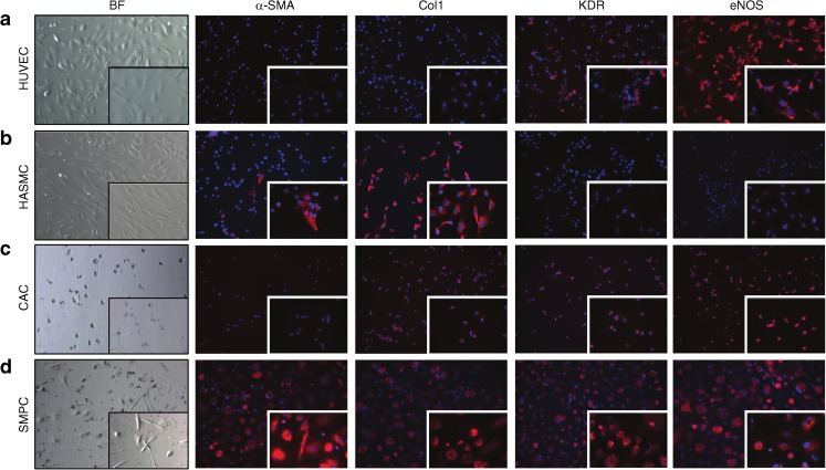
Fig. 3.
The phenotype of in vitro cultured HUVECs, HASMCs, CACs and SMPCs. Pictures were taken at × 200 and × 630 (inset) magnification. (a) HUVECs and (b) HASMCs were used as positive controls for the assessment of expression of EC and SMC differentiation markers. (c) CACs contained collagen type 1, KDR and eNOS, but not α-SMA. (d) SMPCs contained α-SMA, collagen type 1, KDR and eNOS. Nuclear staining is shown in blue (DAPI) while positive staining with the respective antibodies is shown in red. Col1, collagen type 1. BF, Bright field

