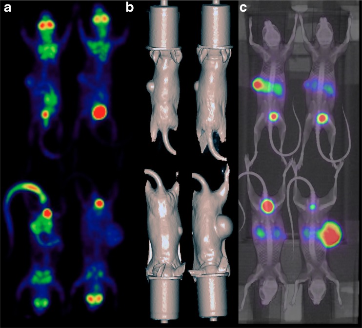
Fig. 5.
Imaging of multiple mice with a positron emitter other than 18F. Four mice with implanted A247 tumours (small axis ranging from 4.6 mm to 12 mm) transfected to overexpress SSTR subclass 2 receptors (cells courtesy of Buck Rogers, Washington University, MI) were imaged 24 h apart on a Mosaic SA-PET system 1.5 h after injection of 18F-FDG (mean activity per mouse 8 ± 0.1 MBq) and 68Ga-DOTATATE (mean activity per mouse 13.2 ± 0.2 MBq). The animals were scanned with a combination of radial (18 mm) and axial (57 mm) displacement. a MIP views for 18F-FDG. b Given the lack of anatomical landmarks on 68Ga-DOTATATE SA-PET images, mice were scanned on a clinical CT scanner and surface images extracted from the CT images. c Fused SA-PET/CT MIP 68Ga-DOTATATE images (SA-PET images are courtesy of David Binns and Carleen Cullinane, Peter MacCallum Cancer Centre, Melbourne, Australia)

