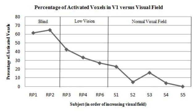
Fig. 4.
Percentage of activated voxels in V1 (FDR < 0.05) versus visual field loss in 5 RP and 5 sighted control subjects. Subjects are ranked along the x-axis in descending order of severity of field loss. Blind subjects were those with minimal light perception and severe tunnel vision, while Low Vision subjects included those with moderate tunnel vision.

