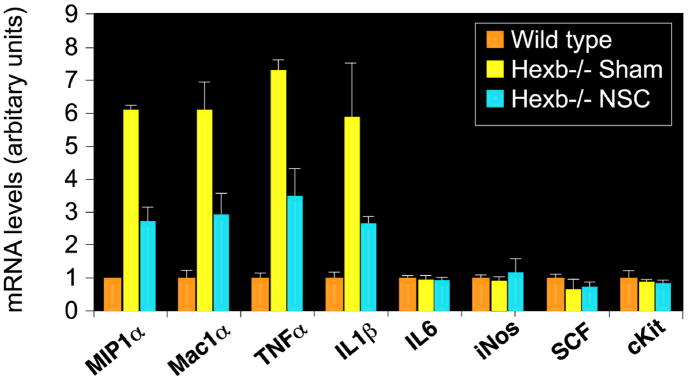
Figure 5. Reduced inflammatory marker expression in brains of NSC-treated Hexb−/− mice.
Expression of inflammatory genes in mouse brains as measured by RT-PCR. The relative gene expression normalized with 18S RNA (arbitrary units) is shown on the graphs. Accompanied by increased levels of microglial/macrophage activation marker Mac1α (Cd11b) (p<0.001), proinflammatory cytokines TNFα and IL-1β and the chemokine MIP1α (CCL3), expression levels were high in the diseased mice, compared to the wild-type mice (p<0.001). Upon NSC treatment, all upregulated genes were reduced significantly when the age-matched sham-treated control group was compared (p<0.001). No significant changes in IL-6, iNOS, SCF or c-kit expression were observed. Data are mean ± s.e.m. based on 3–5 animals in each group (R and L hemispheres averaged together). An unpaired two-tailed Student’s t-test was used to compare data for significance.

