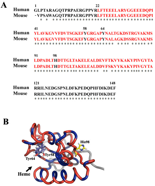
Figure 6.

Comparison of amino acid sequences of GIG47 and NENF and a ribbon diagram of GIG47 showing the potential heme-binding region. (A) Amino acid sequences of GIG47 (human neudesin) and NENF (mouse neudesin) are aligned. The potential heme/steroid binding domain is shown in red. The three residues in bold are the putative heme-binding sites in NENF. (B) A ribbon diagram of GIG47 is shown with three potential heme-binding residues (Tyr58, Tyr64 and His98) displayed in a stick representation mode. The heme-binding domain is colored red. The blue regions in the ribbon indicate hydrophobic residues. A potential heme/steroid binding hydrophobic pocket is clearly visible (indicated by an arrow) between two α-helices (α2 and α3).
