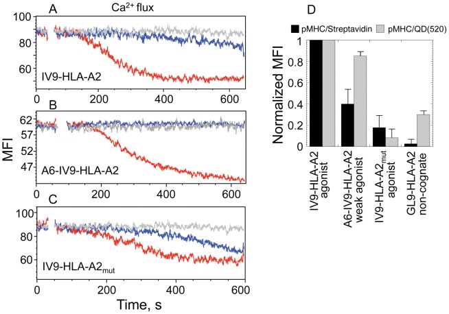
Figure 4. Difference in TCR-mediated signaling kinetics induced by pMHC/QD and pMHC/Streptavidin oligomers.
A, B, C. Time-dependent changes in intracellular calcium concentration in CD8+68A62 CTL induced by indicated pHLA-A2 ligands assembled on either QD(520) (red) and Streptavidin (blue) scaffolds. Concentration of the probes in extracellular medium was the following: 1 nM for IV9-HLA-A2/QD(520) and IV9-HLA-A2/Streptavidin (A), 10 nM for A6-IV9-HLA-A2/QD(520) and A6-IV9-HLA-A2/Streptavidin (B), 5 nM for IV9-HLA-A2mut/QD(520) and IV9-HLA-A2mut/Streptavidin (C). Representative results are shown. D. Relative equilibrium binding of indicated pHLA-A2 ligands assembled on either QD(520) and Streptavidin scaffolds to 62A68 CD8+ CTL is shown. The relative amounts of cell-bound ligands were calculated from MFI measured by flow cytometry. Data represent mean ± s.d.

