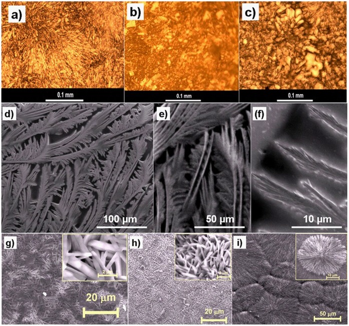
Figure 2. Representative optical microscopy and SEM images of various active pharmaceutical ingredients grown on different substrates synthesised by TE process (a–c) optical images of various Pilocarpine-HCl micro- or nanostrucured morphologies on different substrates like glass (a), polymer foil (b), Ti (c), d–f: SEM images of Ascorbic acid deposited on Si substrate at lower and higher magnifications; g–i: SEM images of Tetracaine-HCl nanostructures deposited on titanium substrate (g), silicon wafer coated with 20 nm Au thin film (h) and silicon coated with 4 nm Au thin film (i).
The inset images inside figures 2 g) to 2 h) show their magnified SEM view of deposited drug respectively.

