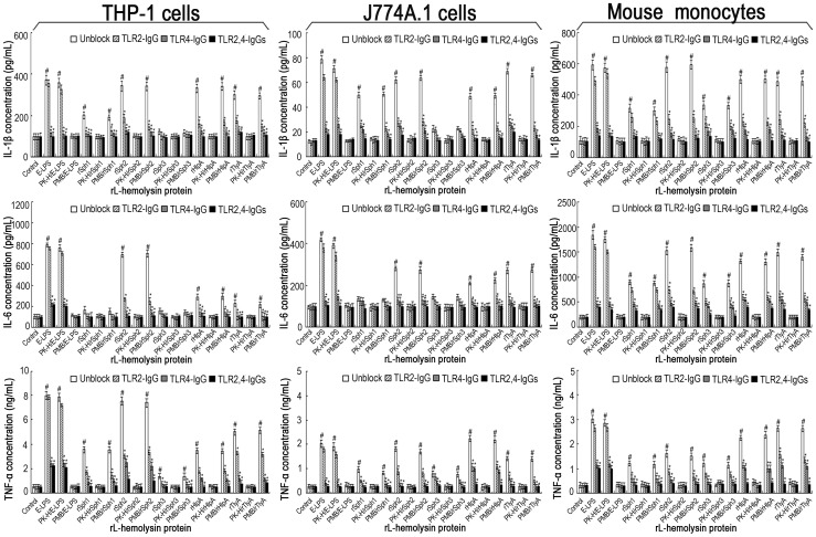
Figure 5. Blocking effects of TLR-IgGs and TLR2 or TLR4 deficieincy on the production of IL-1β, IL-6 and TNF-α under induction of the rL-hemolysins for 24 h.
Bars show the mean± SD of three independent experiments. E-LPS indicates the LPS of E. coli serotype O111:B4. PK-H indicates that the rL-hemolysins or E-LPS were pretreated with proteinase K digestion plus heat–inactivation while PMB indicates that the rL-hemolysins or E-LPS were pretreated with polymyxin B blockade, and were used to monitor possible contamination with E. coli LPS in the rL-hemolysin proteins. The concentration of each of the rL-hemolysins or E-LPS tesed was 1 µg. The mouse monocytes were separated from the peripheral blood samples from TLR2−/−, TLR4−/− or TLR2,4−/− C57BL/6 mice. Control indicates the IL-1β, IL-6 and TNF-α levels in the human THP-1, mouse J774A.1 macrophages, and primary mouse monocytes from wild-type C57BL/6 without TLR2-, TLR4-or TLR2,4-deficience before treatement with any rL-hemolysins or E-LPS. # P<0.05 vs IL-1β, IL-6 and TNF-α levels in the THP-1 or J774A.1 macrophages or primary mouse monocytes before treatment with any rL-hemolysins or E-LPS (control). *P<0.05 vs IL-1β, IL-6 and TNF-α levels in the THP-1 or J774A.1 macrophages that had not been blocked with the TLR2,and/or TLR4 antibodies, or vs the primary monocytes from wild-type C57BL/6 without TLR2-, TLR4-or TLR2,4-deficiency.

