
Figure 8. Subcellular localization of TcOgg1 in T. cruzi.
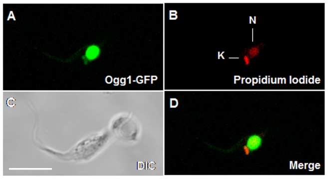
CL Brener strain was transfected with pTREX_TcOGG1, resulting in the expression of TcOgg1 fused with GFP (Ogg1-GFP), which enabled the visualization of the protein under confocal microscope. DNA was stained with propidium iodide. Images obtained were analyzed with Zeiss LSM Image Browser software. N: nucleus; K: kinetoplast (mitochondrion); DIC: differential interference contrast. 5 uM scale bars is present in C.